Case of the Week # XXX Bladder extrophy

Frantisek Grochal
Femicare, Center of prenatal ultrasonographic diagnostics, Martin, Slovakia

Posting Dates: Mar 16, 2026 -

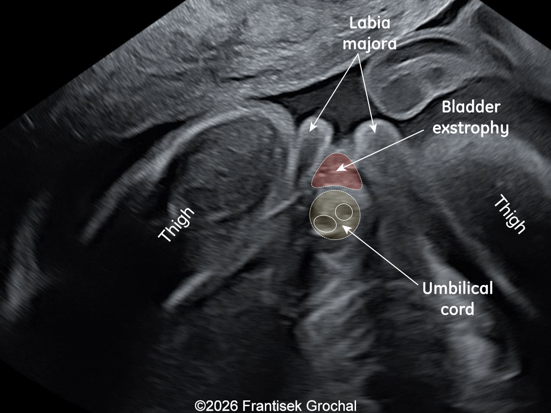
A 26-year-old primigravida with an unremarkable medical history presented for a fetal ultrasound examination at 22+2 weeks’ gestation. The following images and video clips demonstrate the findings identified during the scan.

Image 1
Image 2
Image 3
Video 1 © 2026 Grochal
Video 2 © 2026 Grochal
Video 3 © 2026 Grochal

View the Answer Hide the Answer

Answer

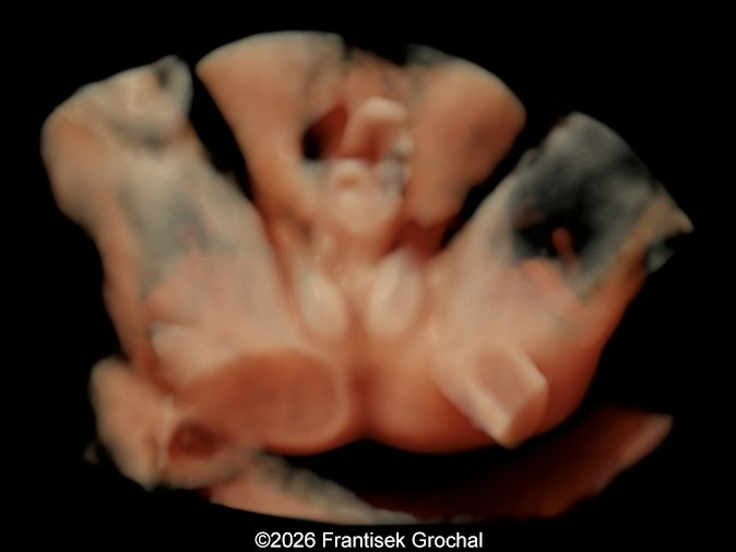
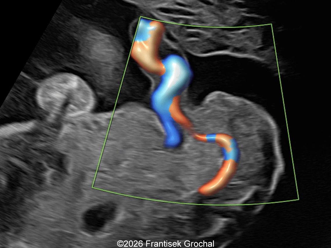
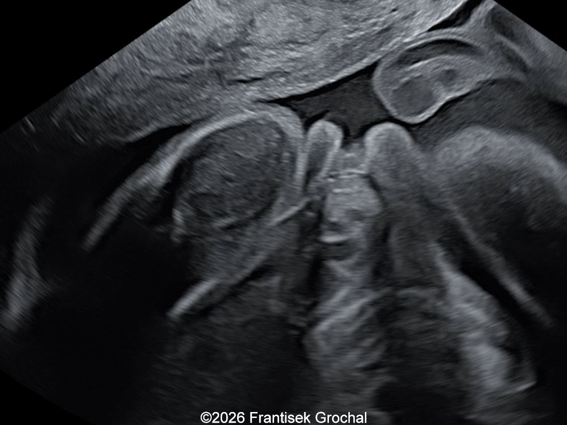
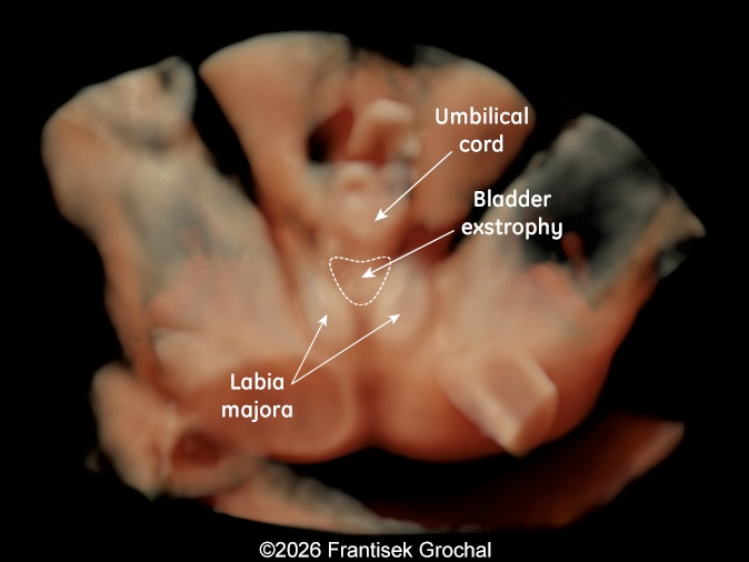
We present a case of bladder extrophy seen in a female fetus at 22+2 weeks’ gestation. The parents opted for the termination of the pregnancy...

3D-representation of the bladder extrophy.
Image 1 3D-representation of the bladder extrophy.
Video 1 Sagittal view: low umbilical cord insertion; urinary bladder not visualized. © 2026 Grochal
Video 2 Both umbilical arteries identified, urinary bladder not visualized. © 2026 Grochal
The image demonstrates a caudal tangential view of the female fetus affected by bladder exstrophy.
Image 2 The image demonstrates a caudal tangential view of the female fetus affected by bladder exstrophy.
Video 3 Normal kideys and amniotic fluid. © 2026 Grochal

Discussion Board

Start a discussion about this case

We appreciate your patience as we review all submitted answers. Check back soon to see if you were correct!

Add to Favorites Favorite

Menu