
Case of the Week (COW)

Current COW
COW Winners
Recently Posted
Current COW
COW Winners
Recently Posted
Current Case of the Week (COW)
Current Case of the Week (COW)
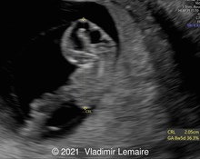
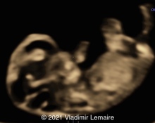
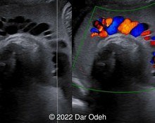
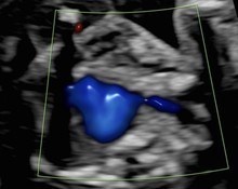
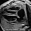
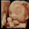
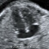
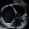
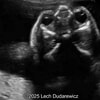
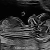
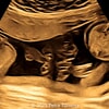
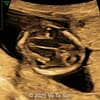
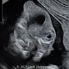
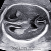
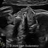
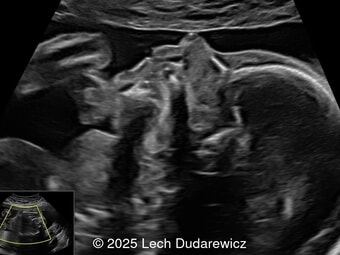
A 33-year-old nullipara with non-contributory medical history presented at 20 weeks, 6 days based on an early scan. Ultrasound revealed the following findings. There were no other apparent abnormalities. What is the most probable diagnosis?
Submit Your Answer
To join the community and participate in solving current cases, simply click the "view current case" button below. This will take you to the case summary page with full details and additional media. To participate, you will first need to create an account or sign-in. You can then submit your answers.
You can only submit your answers once, but you get three answers. The correct answer is revealed at the end of the posting period.
Physicians
Previous Winners
-
Javier Cortejoso, Spain
Cases Solved: 21 -
Annette Reuss, Germany
Cases Solved: 18 -
Ionut Valcea, Romania
Cases Solved: 17 -
Mayank Chowdhury, India
Cases Solved: 16 -
Alexandr Krasnov, Ukraine
Cases Solved: 15
First-Time Winners
-
Ali Ozgur Ersoy, Turkey
Cases Solved: 11 -
ZHANNA Kurmangaliyeva, Kazakhstan
Cases Solved: 11 -
Đặng Mai Quỳnh, Viet Nam
Cases Solved: 9 -
ANA PAULA PASSOS, Brazil
Cases Solved: 9 -
Amparo Gimeno, Spain
Cases Solved: 9
Sonographers
Previous Winners
-
Dianna Heidinger, United States
Cases Solved: 14 -
Kimberly Delaney, United States
Cases Solved: 11 -
Eti Zetounie, Israel
Cases Solved: 9 -
CHERYL TURNER, United States
Cases Solved: 7 -
Padmanaban Koochu Govindaraju, United Kingdom
Cases Solved: 4
First-Time Winners
-

CHEN YANG, China
Cases Solved: 10 -
ASHLEA HARDIN, United States
Cases Solved: 8 -
Fred Pop, Uganda
Cases Solved: 8 -
CARLOS JOSE PIÑA VILLEGAS, Peru
Cases Solved: 5 -
Joanne Maloney, United States
Cases Solved: 5
Contributors
Top Contributors
-
Lech Dudarewicz, Poland
Article & Case Contributions: 9 -
Javier Cortejoso, Spain
Article & Case Contributions: 6 -
Frantisek Grochal, Slovakia
Article & Case Contributions: 3 -
Vladimir Lemaire, United States
Article & Case Contributions: 3 -
Petra Turnova, Slovakia
Article & Case Contributions: 2
Previous winners: Users who have been recognized on a "Top Winners list" in years past; First-time winners: Users who have not yet been on the annual "Top Winners list." The Sonographers category also incorporates "other" job titles.
We Would Like Your Feedback
Which was your favorite case of the week in 2025? Your favorite case may be the one with the most demonstrative images and videos, a particularly well-written and informative discussion, or an unusual and unique disease process.
News & Notes
Dear Esteemed Users of TheFetus.net,
We had 22 cases of the week this year with unique and complex conditions that varied from Torcular Herophili Thrombosis to Prune Belly Syndrome to Sternoschisis. We want to hear which one was your favorite!
Your favorite case may be the one with the most demonstrative images and videos, a particularly well-written and informative discussion, or an unusual and unique disease process. To vote for your favorite case, log into… read the full entry
TheFetus.net
© 2014 - 2025 TheFetus.net