Meckel-Gruber syndrome

Omayyah Dar-Odeh (1), Samih Abdel-Jawad (1) , Rua'a Abdel-jawad (2), Raghad Abdel-Jawad (3), Rahaf Abdel-Jawad (3)
(1) Private sector; (2) King Husein Medical center; (3) Hashemite University

Posting Dates: Oct 25, 2024 - Oct 30, 2024

35-year-old G7P6 woman involved in a consanguineous marriage with a maternal relative was referred at 19 weeks, 2 days for detailed scan because of an obstetric history significant for two miscarriages, and both a male and female newborn delivered with congenital anomalies who died soon after birth. The couple had a healthy girl and boy. Our prenatal imaging showed the following findings:

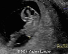
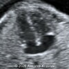
Image 1
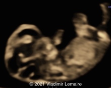
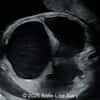
Image 2
Video 1 © 2024 Omayyah Dar-Odeh
Video 2 © 2024 Omayyah Dar-Odeh
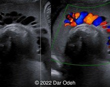
Video 3 © 2024 Omayyah Dar-Odeh
Video 4 © 2024 Omayyah Dar-Odeh
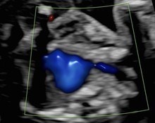
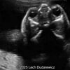
Image 3

View the Answer Hide the Answer

Answer

We present a case of Meckel Gruber syndrome.

Our imaging, which was limited because of maternal body habitus (high BMI) and oligohydramnios, showed:

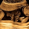
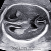
  • Image 1: Transverse section in abdomen showing bilateral enlarged cystic kidneys
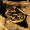
  • Image 2: Postaxial polydactyly in foot
  • Video 1: Postaxial polydactyly in hand
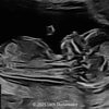
  • Video 2: Abnormal skull shape and occipital encephalocele
  • Video 3: Enlarged cystic kidneys
  • Video 4: Club foot
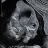
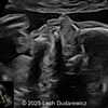
  • Image 3: Large abdominal circumference

We suspected Meckel Gruber syndrome and advised the patient to consider amniocentesis for genetic study.  Amniocentesis was delayed due to poor patient compliance and was technically difficult due to anhydramnios. Therefore, chorionic villous sampling was done.

Whole exome sequencing demonstrated a homozygous c.513C>G / p. Tyr171Ter class 2 variant in the B9D2 gene. This result is expected to lead to B9D2 Related Disorder Phenotype (OMIM: 611951). B9D2 (B9 Domain Containing 2) is a protein coding gene and diseases associated with B9D2 include Meckel Syndrome, Type 10 and Joubert Syndrome 1.  The patient was counselled regarding the lethal outcome. Genetic screening and clinical evaluation in family members were recommended in order to determine the pathogenic effect of this change.

The couple refused termination and continued follow up at a peripheral public hospital.  The patient continued the pregnancy until 38 weeks and underwent lower uterine segment cesarean section because of obstetric reason (three prior cesarean sections) at a public hospital.  A live male fetus weighing 2400 grams with multiple anomalies (microcephaly, large occipital encephalocele, distended abdomen, postaxial polydactyly in hands and feet) was delivered. Soon after birth, the newborn had difficulty breathing with cyanosis and died 3 hours later. Autopsy was denied by the parents.

The newborn with occipital encephalocele
Image 1 The newborn with occipital encephalocele
Postaxial polydactyly in foot
Image 2 Postaxial polydactyly in foot
Postaxial polydactyly in hand and misshaped ear
Image 3 Postaxial polydactyly in hand and misshaped ear
The newborn with microcephaly, postaxial polydactyly in hands and feet
Image 4 The newborn with microcephaly, postaxial polydactyly in hands and feet

Discussion

Meckel–Gruber syndrome (MKS) is a rare, lethal and autosomal recessive disease with a 25% recurrence rate [1,2]. It is caused by mutations in genes encoding proteins that are structural or functional components of the primary cilium. Conditions that are caused by mutations in ciliary genes are collectively termed the ciliopathies, and MKS represents the most severe condition in this group of disorders [14].

It was originally described by Johann Friedrich Meckel in 1822. G.B. [6,12]. Gruber also published reports of patients with MKS in 1934 and gave it the name dysencephalia splanchnocystica. Worldwide, its incidence is 1 per 13,250–140,000 livebirths [5,10]. Higher incidences are observed in endogamous populations, such as Gujarati Indians, Tatars, Hutterites, and in Finland where 1 in every 9,000 live births is affected [14]. An increased incidence has been reported in regions where consanguineous marriages are common (with 1 affected birth per 1,300).

It is characterized by the triad of cystic renal disease, a central nervous system malformation, and polydactyly  [7,8,9]. The most common findings include renal cystic dysplasia (95-100%), occipital encephalocele (60-80%), and postaxial polydactyly (55-70%) [16].  At least two of the major antenatal ultrasound findings should be present for diagnosis of this syndrome. In approximately 57% of the cases, the three findings can be seen together. In addition to these classical findings, central nervous system findings can also include Dandy-Walker malformation, Arnold Chiari malformation, microcephaly, and hydrocephalus. Other anomalies include eye defects, cleft palate/lip, hepatic duct malformation characterized by proliferation and fibrosis in the portal areas of the liver and bile ducts, pancreatic cysts and fibrosis, congenital heart disease, adrenal hypoplasia, and genitourinary malformations such as male genital organ hypoplasia, male pseudohermaphroditism, cryptorchidism, ureter agenesis, hypoplasia or duplication, and bladder agenesis and hypoplasia. Characteristic images on ultrasound are dependent on gestational age. While the classical triad can be better identified by ultrasound before the 14th week of pregnancy, it becomes difficult to diagnose with advanced oligohydramnios as gestation progresses.

Meckel–Gruber syndrome has extreme genetic heterogeneity and displays allelism with other ciliopathies such as Joubert syndrome (JBTS), COACH syndrome (cerebellar vermis hypo/aplasia, oligophrenia, congenital ataxia, ocular coloboma, and hepatic fibrosis), oro-facio-digital syndrome (OFD), nephronophthisis (NPHP), and Bardet–Biedl syndrome (BBS) [15]. To date, mutations in 14 genes are identified as causative for MKS. Mutations in a further three genes, are implicated on the basis of pathogenic private mutations in individual families with either MKS or MKS-like phenotypes. Two other genes, comprising SEC8 (also known as EXOC4) and EXOC3L2, have private mutations as a cause of probable MKS phenotypes [4]. In total, mutations in these genes appear to explain only 50–60% of MKS cases [14].  Most of these genes are also responsible for Joubert syndrome, leading to the concept that MKS is the extreme lethal phenotype of Joubert syndrome [16].

Genetic mutations on chromosome 17q21-24 were determined in most of the cases. Pathogenesis of MGS is related to dysfunction of primary cilia. Because of the elevated genetic heterogeneity potential, mapping is required on the other different locations. Additionally new genes accompanied with this syndrome were discovered and that explains phenotypical expression of this syndrome [11].

Trisomy 13 and Smith Lemli-Opitz syndrome should be denied in the differential diagnosis in cases including multiple anomalies (CNS, cardiac, renal and extremity) [1].

The prognosis in Meckel-Gruber syndrome is poor and it is a lethal condition owing to dysplastic kidneys that result in severe oligohydramnios and pulmonary hypoplasia [16].

References

[1] Erol SA, Kırbaş A. Prenatal Sonographic Diagnosis of Meckel-Gruber Syndrome: A Case Report. Med J West Black Sea. 2021;5(1):110-113.
[2] Raj M, Dhanuka S, Agarwal P, et al. Meckel Gruber syndrome – a case report. Surg Exp Pathol. 2020 April 15:3:11.
[3] Srivastava, S, Manisha R, Dwivedi A, et al. Meckel Gruber and Joubert Syndrome Diagnosed Prenatally: Allelism between the Two Ciliopathies, Complexities of Mutation Types and Digenic Inheritance.  Fetal Pediatr Pathol. 2022 Dec;41(6):1041-1051.
[4] Stembalska A, Rydzanicz M, Pollak A, et al. Prenatal Versus Postnatal Diagnosis of Meckel-Gruber and Joubert Syndrome in Patients with TMEM67 Mutations. Genes (Basel). 2021 Jul;12(7):1078.
[5] Aslan K, Aslan EK, Orhan A, et al. Meckel Gruber syndrome, A case report. Organogenesis. 2015 Jun;11(2):87-92.
[6] No Author. Johann Friedrich Meckel, the Younger (1781-1833). JAMA. 1970 Oct 5;214(1):138-9.
[7] Gazioğlu N, Vural M, Seçkin MS, et al. Meckel-Gruber syndrome. Childs Nerv Syst. 1998 Mar;14(3):142-5.
[8] Reis I, Ferreira P, Saraiva S, et al. VP04.20: Meckel–Gruber syndrome: the role of early ultrasound – a case report. Ultrasound Obstet Gynecol. 2020 Oct; 56(S1):72.
[9] Sergi C, Adam S, Otto HF, et al. Study of the Malformation of Ductal Plate of the Liver in Meckel Syndrome and Review of Other Syndromes Presenting with this Anomaly. Pediatr Develop Pathol. 2000 Nov;3(6):568-583.
[10] Siraj R, Rehan R, Nasir S, et al. The Exceptional Case of Healthy Precious Infant Surviving with Meckel-Gruber Syndrome Variant. Med Case Report. 2021 Dec;7(11):211.
[11] Darouich SI, Bettaieb M, Khila T, et al. Phenotypic variability in Meckel-Gruber syndrome. Cilia. 2015 Jul;4(Suppl 1):P1.
[12] Bissonnette B, Luginbuehl I, Engelhardt T. "Meckel-Gruber Syndrome." Syndromes: Rapid Recognition and Perioperative Implications, 2nd edition. New York: McGraw Hill, 2019.
[13] Hartill V, Szymanska K, Sharif SM, et al. Meckel-Gruber Syndrome: An Update on Diagnosis, Clinical Management, and Research Advances. Front Pediatr. 2017 Nov 20:5:244.
[14] Shetty BP, Alva N, Patil S, et al. Meckel-Gruber Syndrome (Dysencephalia Splanchnocystica). J Contemp Dental Pract 2012 Sep;13(5):713-715.
[15] Kheir AEM, Imam A, Omer IM, et al. Meckel-Gruber syndrome: A rare and lethal anomaly. Sudan J Paediatr. 2012; 12(1): 93–96.
[16] Roy J, Pal M. Meckel Gruber Syndrome. J Clin Diagn Res. 2013 Sept;7(9):2102-2103.

Discussion Board

Join the discussion about this case

We appreciate your patience as we review all submitted answers. Check back soon to see if you were correct!

Add to Favorites Favorite

Menu