Case of the Week #632
UT Southwestern Medical Center, Plano, Texas, United States of America
A 39-year-old patient, G1P0, presented to our maternal fetal medicine unit at 30 weeks of gestation for a growth scan. The fetus was male with low-risk noninvasive prenatal testing. The following findings were observed.
View the Answer Hide the Answer
Answer
We present an isolated case of Tetralogy of Fallot (TOF). The diagnosis was confirmed by pediatric cardiology.
Our images demonstrate the following:
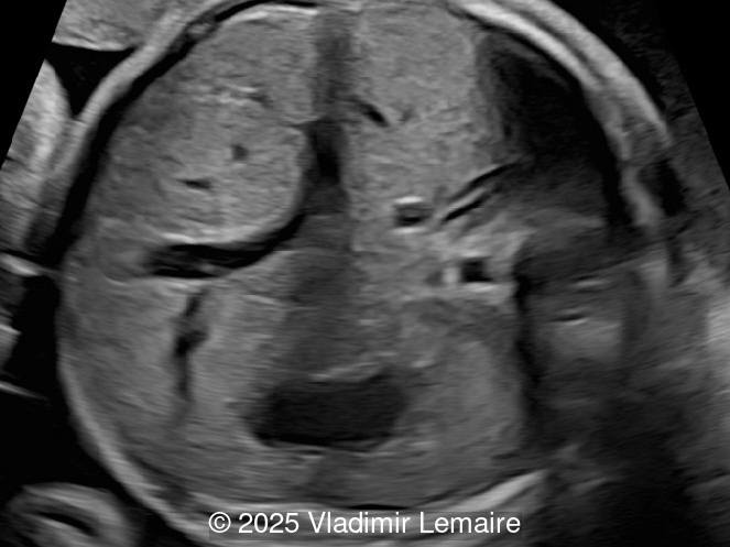
- Image 1: Transverse view of the upper abdomen at the level of the abdominal circumference.
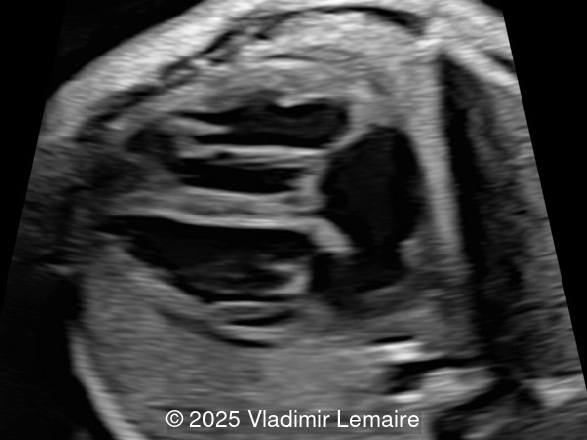
- Image 2: Axial plane of the fetal chest at the level of the four-chamber view. Note the normal appearance of the four-chamber view.
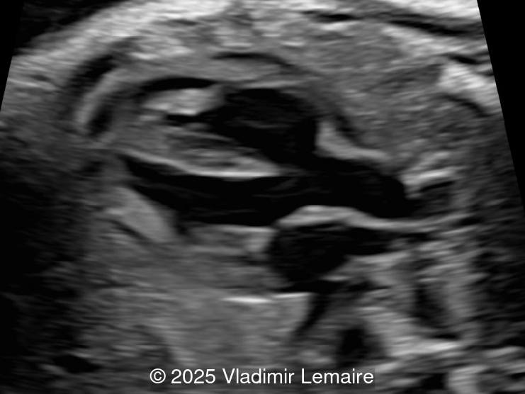
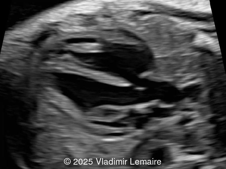
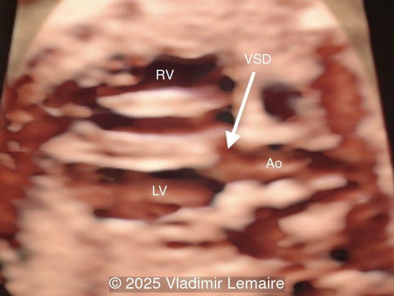
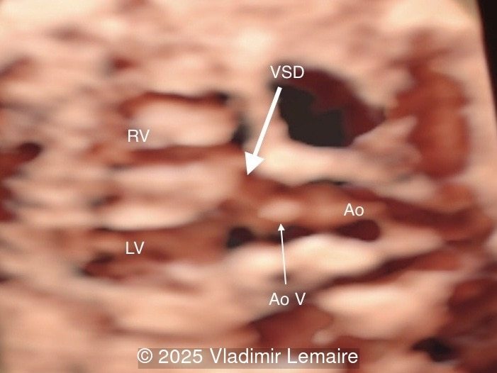
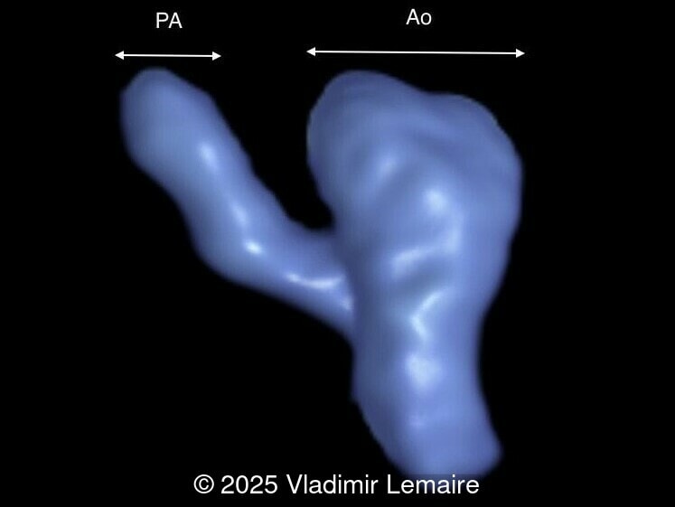
- Images 3 and 4: The five-chamber view shows the ventricular septal defect and the dilated overriding aorta.
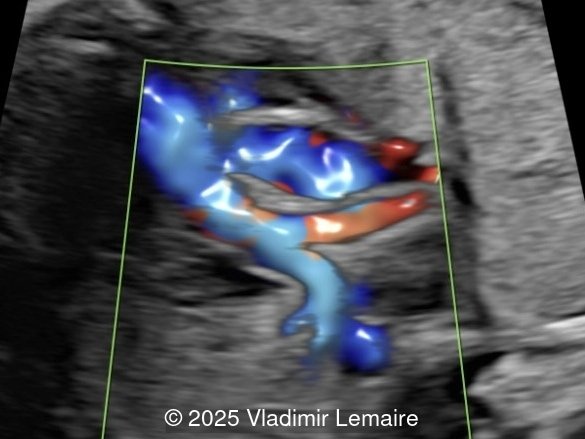
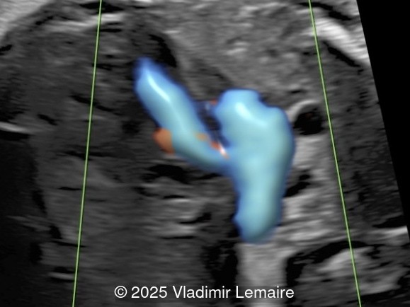
- Image 5: The right ventricular outflow tract view with color Doppler demonstrates a small main pulmonary artery when compared to the dilated aorta due to pulmonary stenosis.
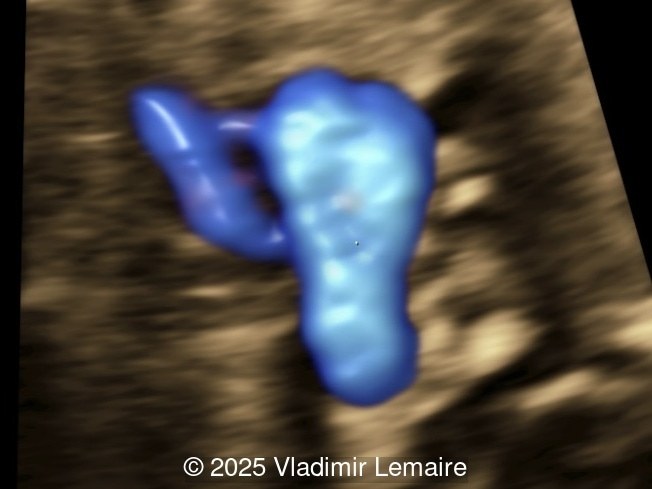
- Image 6: The three-vessel-trachea view with color Doppler shows the discrepant size of the great arteries with the pulmonary artery smaller than the aorta. Note the presence of antegrade blood flow in both arteries.
In the images below, the following abbreviations are used: RV (right ventricle); LV (left ventricle); Ao (Aorta), VSD (ventricular septal defect), Ao V (Aortic valve); PA (pulmonary artery)
Discussion
Tetralogy of Fallot (TOF) is characterized by a malaligned ventricular septal defect (VSD), an aortic root that overrides the ventricular septal defect, and an infundibular pulmonary stenosis. Right ventricular hypertrophy, the fourth anatomic feature of the tetralogy, is not present prenatally. Tetralogy of Fallot, one of the most common forms of cyanotic congenital heart disease, is found in about 1 in 3,600 live births and accounts for 3 to 7% of infants with congenital heart disease. There are several variations of Tetralogy of Fallot, the most common of which is with pulmonary stenosis and accounts for about 80% of all newborns with the condition. The spectrum of disease also includes severe forms such as TOF with pulmonary atresia and TOF with absent pulmonary valve.
In Tetralogy of Fallot, the four-chamber view typically appears normal unless a large ventricular septal defect is visible in this plane. TOF is usually detected in the left ventricular outflow tract view, which demonstrates a perimembranous, subaortic ventricular septal defect with an overriding aorta. The overriding aorta is due to the discontinuity between the interventricular septum and medial wall of the aorta (malalignment VSD). This results in a partial connection of the aorta to the right ventricle, with an aorta that is slightly shifted to the right, referred to as aortic dextroposition. The aortic root appears dilated, especially in the third trimester, because it receives blood from both the right and left ventricles. The overriding aorta has a parallel course to the interventricular septum in contrast to the ascending aorta in a normal heart.
The demonstration of a narrow but patent main pulmonary artery is also required for the diagnosis of Tetralogy of Fallot. This is best demonstrated at the level of the short-axis or the three-vessel view. The discrepancy between the pulmonary trunk and the aorta can be subtle in midgestation, however it becomes more obvious with advancing gestation.
Color Doppler confirms the presence of an overriding aorta with blood draining from both ventricles, through the ventricular septal defect into the aortic root. Due to high perfusion, inflow into the aorta appears aliased. At the level of the three-vessel-trachea view, color Doppler can also demonstrate a small pulmonary artery. Flow is antegrade across the ductus arteriosus in mild Tetralogy of Fallot and can be reversed in severe cases. Color Doppler can help differentiate various subgroups of TOF as postnatal ductal dependency of the pulmonary circulation can be associated with cyanosis of the newborn.
On prenatal ultrasound, associated cardiac abnormalities include a right aortic arch in 25% of cases and occasionally an atrioventricular septal defect which increases the risk of chromosomal abnormalities. A patent foramen ovale or an atrial septal defect and a persistent left superior vena cava have been reported in 83% and 11%, respectively, of newborns with Tetralogy of Fallot.
Associated extracardiac anomalies are common. Chromosomal abnormalities, mainly trisomies 21, 13 and 18, are found in 30% of cases. Deletion 22q11.2 occurs in 10 to 15% of fetuses and newborns with Tetralogy of Fallot, and is more likely in cases with a hypoplastic thymus, right-sided aortic arch, presence of extracardiac anomalies, or polyhydramnios. TOF can also be associated with syndromic conditions such as Alagille syndrome and CHARGE syndrome.
Differential diagnosis includes pulmonary atresia with ventricular septal defect, absent pulmonary valve syndrome, common arterial trunk, double outlet right ventricle, and malaligned VSD without abnormalities of the great vessels. All of these conditions present with an overriding aorta.
Pulmonary artery growth has been shown to be variable and unpredictable. Therefore, serial prenatal ultrasound examinations to assess fetal pulmonary artery growth and flow across the ductus arteriosus are crucial for counseling and appropriate care of the newborn. Findings that have been associated with poor prognosis include decelerated growth of the pulmonary artery, accelerated growth of the ascending aorta and cessation of forward flow through the pulmonic valve with reversed flow through the ductus arteriosus. Pulmonary atresia with ventricular septal defect and absent pulmonary valve syndrome are known to have a worse prognosis.
References
Abuhamad, A, et al. "Tetralogy of Fallot". A Practical Guide to Fetal Echocardiography: Normal and Abnormal Hearts (4th edition). Singapore: Wolters Kluwer, 2022. pgs 410-425.
Discussion Board
Winners

Dianna Heidinger United States Sonographer

Javier Cortejoso Spain Physician

paola quaresima Italy Physician

Andrii Averianov Ukraine Physician

Alexandr Krasnov Ukraine Physician

Andres Arencibia United States Physician

Mayank Chowdhury India Physician

Olha Petrenko Ukraine Physician

Adrian Popa Romania Physician

Nutan Thakur India Physician

Shilpen Gondalia India Physician

Boujemaa Oueslati Tunisia Physician

Tatiana Koipish Belarus Physician

carlos lopez Venezuela Physician

Aysegul Ozel Turkey Physician

Caroline Reichert Garcia Brazil Physician

Alvaro Gómez Mexico Physician

Anita Silber Israel Physician
Viorel Suciu-Lazar United States

Kimberly Delaney United States Sonographer

Marianovella Narcisi Italy Physician
Javier Ayala Spain Physician

Rebecca Evans Australia Sonographer

CHEN YANG China Physician

Amparo Gimeno Spain Physician

Elena Andreeva Russian Federation Physician
Patrícia Silva Portugal Physician
Basem Hamed United States

Muradiye YILDIRIM Turkey Physician

Ta Son Vo Viet Nam Physician

ALBANA CEREKJA Italy Physician

Eti Zetounie Israel Sonographer

SAMUEL GELVEZ TELLEZ Colombia Physician

Murat Cagan Turkey Physician
Umutcan KAYIKÇI Turkey Physician

ANA PAULA PASSOS Brazil Physician

Büşra Cambaztepe Turkey Physician
Gayane Begjanyan Armenia Physician

Ionut Valcea Romania Physician

Đặng Mai Quỳnh Viet Nam Physician
Halil Korkut Dağlar United States Physician

Hien Nguyen Van Viet Nam Physician

Almaz Kinzyabulatov Russian Federation Physician

Kareem Haloub Australia Physician

Zuzana Briešková Slovakia Physician
Katarína Pitoňáková Czech Republic Physician
András Weidner Hungary Physician
Maria Kuznetsova Russian Federation Physician

Thomson Thomson Indonesia OB-GYN

Fred Pop Uganda Sonographer

Annette Reuss Germany Physician

Arati Appinabhavi India Physician

shruti Agarwal India Physician

Jay Vaishnav India Physician

CHERYL TURNER United States Sonographer

YULIA VISHNEVSKAYA Russian Federation Physician
Hayley Jacobson Israel Physician

Nguyen Xuan Cong Viet Nam Physician
Perrine Riou-Kerangal French Polynesia Sage-femme échographiste

Lynn Davis United States Sonographer

Sruthi Pydi India Physician
Laura Wharton United Kingdom Physician
Apoorva Mannikar India Physician
Rohit Sanghani India Physician
Rupal Sasani India Physician

philip pattyn Belgium Physician
Joanna Głowska-Ciemny Poland Physician
Duc Giang Viet Nam Physician

Petra Barboríková Slovakia Physician
Dolly Agrawal India Physician
Rajnikant Vasava India Physician

Denys Saitarly Israel Physician

Le Tien Dung Viet Nam Physician

Tetiana Ishchenko Ukraine Physician
Costin Radu Lucian Romania Physician
Julia Knypinski United States Physician
Zina Kerbi Algeria Physician
Molly Brown United States Sonographer
María Victoria Peral Parrado Spain Physician

Le Duc Viet Nam Physician
Janaina Andrade Brazil Physician
Mahesh Kilje India Physician

Hana Habanova Slovakia Physician
Kelsey O'Brien United States Sonographer
Tushar verma India Physician
Jesus Cortez Venezuela Physician

Gnanasekar Periyasamy India Physician
Molly Paulson United States Sonographer
Maureen Cosentino United States Sonographer
Navya Sri Mopada India Physician
Rinku Vasaya Congo, The Democratic Republic of The Sonographer

Petra Tallova Slovakia Physician
José Velasco Ecuador Physician
Tal Lesser United States Physician
Diogo Santos Portugal Physician
Valerie Finkelstein United States Sonographer

Kurmanzhan Balmukhambetova Kazakhstan Physician

Ali Ozgur Ersoy Turkey Physician
Ayse Ceren Duymus Turkey Physician
Lucia Bobik Slovakia Physician
Karla Eutsler United States Sonographer

Manuel Rodriguez Mexico Physician

ZHANNA Kurmangaliyeva Kazakhstan Physician
Liam McCullough Slovakia Physician
Dubyanskaya Yuliya Russian Federation Physician
Albert Guarque Rus Spain Physician

KIM SOCHETRA Cambodia Physician
Henrietta Karlsson Spain Physician
P K Indonesia Physician

Dang Thinh Nguyen Viet Nam Physician
Jose Carcamo Honduras Physician
Kristina Gonosova Slovakia Sonographer
Khanh Dinh Le Tran Viet Nam Physician
jimena salcedo Spain Physician
Kim Wong Mexico Physician
Ramz Rafiq Australia Sonographer
Neelesh Patel India Physician