Case of the Week # 589
(1) Professor of Obstetrics & Gynecology, Haifa, Israel; (2) St Mary's Medical Center, San Francisco, CA, USA
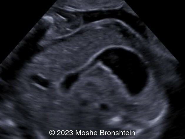
A patient presents at 14 weeks gestation with the following findings.
View the Answer Hide the Answer
Answer
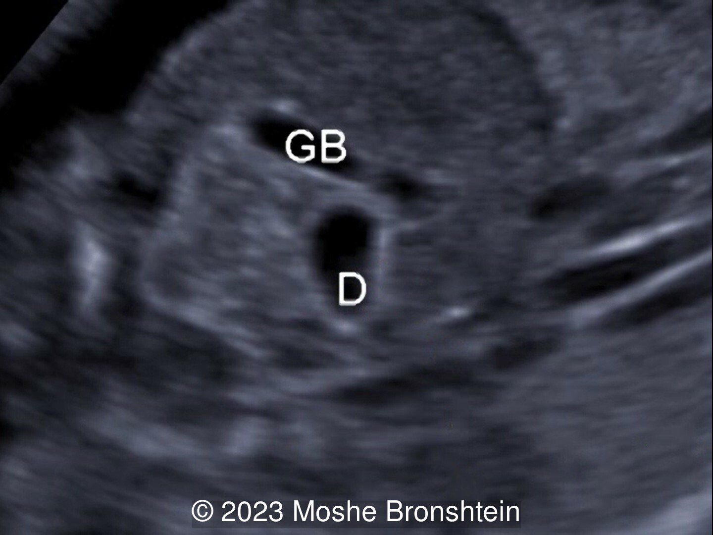
We present a case of duodenal atresia involving the fourth portion of the duodenum.
Ultrasound examination at 14 weeks and confirmed at 19 weeks demonstrated a transverse image of fetal abdomen with stomach on the left side (bigger bubble of so called “double bubble sign”) connected via thinner part of duodenum to slightly dilated distal part of duodenum (second bubble of so called “double bubble sign”). Not shown is an aberrant right subclavian artery and absent ductus venosus. Chromosome analysis, including microarray and exome sequencing, were normal. The fetus developed mild polyhydramnios at 27 weeks gestation, which progressed to severe polyhydramnios by 34 weeks.
Surgical exploration at birth revealed duodenal atresia involving the 4th portion of the duodenum. There was no intestinal malrotation identified at surgery.
Discussion
Duodenal atresia has an incidence of 1 per 5000–10,000 live births. Intrinsic duodenal atresia and stenosis most commonly occur in the first and second portions of the duodenum [1]. Those found in the third and fourth portion of the duodenum can be associated with malrotation and midgut volvulus. Ladd’s bands, caused by incomplete absorption of cecal and ascending colon mesenteries, can cause external compression of the duodenum and be associated with malrotation [1]. Duodenal obstruction can be classified as complete and incomplete. Complete duodenal obstruction usually results from bowel atresia or annular pancreas, whereas incomplete atresia can be caused by an intraluminal web [2].
Prenatal detection rates of duodenal obstruction vary between 33-48% [2,3]. In patients with complete obstruction, the rate of diagnosis before birth may be up to 73% [2]. In a study reviewing 33 cases of duodenal obstruction, no infants with incomplete obstructions were diagnosed prenatally [2]. Prenatal diagnosis of duodenal obstruction occurs at a median age of 25 weeks [2], with rare reports of early diagnosis at 14-15 weeks [4, 5].
Findings on ultrasound include a double bubble from dilation of the stomach and proximal duodenum. The differential diagnosis for a double bubble includes normal peristalsis [5], choledochal cyst [6], or duodenal duplication [7]. Establishing intestinal continuity can differentiate between duodenal atresia and choledochal cyst or duodenal duplication [3]. Additionally, fetuses with duodenal atresia can present with polyhydramnios, though the rate was only 33% in patients with complete obstruction and patients with incomplete obstruction had normal amounts of amniotic fluid [2]. Fetuses with more than two bubbles have been reported and can be found to have a combination of pathologies including malrotation and annular pancreas [8]. Evaluation by MRI [8] or 3D ultrasound [9] can further delineate the anatomy.
Duodenal atresia is often associated with other anomalies. In a study reviewing 61 cases of duodenal atresia, only 43% were isolated [3]. Associated anomalies included Down’s syndrome (46%), cardiac anomalies (31%), malrotation (10%), bowel atresia (7%), and VACTERL association (vertebral, anorectal, tracheoesophageal, renal, limb) (5%) [3]. In a review of the literature by Kimble et al, 29% of infants with duodenal atresia had malrotation. The authors hypothesize that some cases of duodenal atresia and stenosis may occur due to compression of the embryonic duodenum by crossing bands from a malrotated colon [10].
Our case is unique in that the diagnosis was made early in gestation at 14 weeks, and that the location of the stenosis occurred in the 4th portion of the duodenum without associated malrotation.
References
[1] Russo P, Huff D. “CHAPTER 8 - Congenital and Developmental Disorders of the GI Tract.” Surgical Pathology of the GI Tract, Liver, Biliary Tract, and Pancreas (Second Edition). W.B. Saunders, 2009. pg 145-168.
[2] Saalabian K, Friedmacher F, Theilen TM, et al. Prenatal Detection of Congenital Duodenal Obstruction-Impact on Postnatal Care. Children (Basel). 2022 Jan 26;9(2):160.
[3] Choudhry MS, Rahman N, Boyd P, et al. Duodenal atresia: associated anomalies, prenatal diagnosis and outcome. Pediatr Surg Int. 2009 Aug;25(8):727-30.
[4] Petrikovsky BM. First-trimester diagnosis of duodenal atresia. Am J Obstet Gynecol. 1994 Aug;171(2):569-70.
[5] Zimmer EZ, Bronshtein M. Early diagnosis of duodenal atresia and possible sonographic pitfalls. Prenat Diagn. 1996 Jun;16(6):564-6.
[6] Dewbury KC, Aluwihare AP, Birch SJ, et al. Prenatal ultrasound demonstration of a choledochal cyst. Br J Radiol. 1980 Sep;53(633):906-7.
[7] Malone FD, Crombleholme TM, Nores JA, et al. Pitfalls of the 'double bubble' sign: a case of congenital duodenal duplication. Fetal Diagn Ther. 1997 Sep-Oct;12(5):298-300.
[8] Hung JH, Shen SH, Chin TW, et al. Prenatal diagnosis of double duodenal atresia by ultrasound and magnetic resonance image. Prenat Diagn. 2007 Apr;27(4):381-3.
[9] López Ramón Y Cajal C, Ocampo Martínez R. Prenatal diagnosis of duodenal atresia with three-dimensional sonography. Ultrasound Obstet Gynecol. 2003 Dec;22(6):656-7.
[10] Kimble RM, Harding J, Kolbe A. Is malrotation significant in the pathogenesis of duodenal atresia? Pediatr Surg Int. 1995 Jul;10:325-28.
Discussion Board
Winners
Emad Abdelrahim Elshorbagy Egypt Physician
Dianna Heidinger United States Sonographer
Javier Cortejoso Spain Physician

Padmanaban Koochu Govindaraju United Kingdom Sonographer
Kristína Bihariová Slovakia Physician
Igor Yarchuk United States Sonographer
Iuliia Iudina Russian Federation Physician

belen garrido Spain Physician

Dr.Neel Vaghasia India Physician
Ana Ferrero Spain Physician
Mayank Chowdhury India Physician
Nutan Thakur India Physician

Oskar Sylwestrzak Poland Physician
Vladimir Lemaire United States Physician

Shilpen Gondalia India Physician

DAVID BEAUMONT United Kingdom Physician
Ivan Ivanov Russian Federation Physician

Boujemaa Oueslati Tunisia Physician
Sara Abdallah Salem Egypt Physician

Wale Adediran United States Sonographer

Michal Michna Slovakia Physician
Halil Mesut Turkey Physician
Rushina Patel United States Sonographer
Emiliano Ciari Argentina Physician

Philippe Deblieck Germany Physician

Peter conner Sweden Physician
Khatuna Avaliani United States Physician
Kimberly Delaney United States Sonographer

Olivia Ionescu United Kingdom Physician

Kirill Suprutskii Russian Federation Physician
Marianovella Narcisi Italy Physician

Anna Marin Russian Federation Physician
Suat İnce Turkey Physician

Faten Badr Egypt Physician
Shari Morgan United States Sonographer
Liem Dang Le Viet Nam Physician

Amparo Gimeno Spain Physician
Anna Kalinina Russian Federation Physician
Muradiye YILDIRIM Turkey Physician

ALBANA CEREKJA Italy Physician

Yana Semina Russian Federation Physician
Eti Zetounie Israel Sonographer
Murat Cagan Turkey Physician
Eslam Adel ammar Egypt Physician
Loai Said Palestinian Territory, Occupied Physician
Rasha Abo Almagd Egypt Physician

Ionut Valcea Romania Physician
Đặng Mai Quỳnh Viet Nam Physician

mevlüt bucak Turkey Physician

Halil Korkut Dağlar United States Physician

Karin Tinnemeier Germany Physician

Borisova Elena Russian Federation Physician
Almaz Kinzyabulatov Russian Federation Physician

zhang jie China Physician

Kareem Haloub Australia Physician

carlos lugo-leon Venezuela Physician

Dr Monika Sharma India Physician
Martina Vagaská Slovakia Physician

Laura Luse Latvia Physician
Manar Osman Egypt Physician

Vasanth Kumar India Physician
Arati Appinabhavi India Physician
BHOOMIKA SHARMA India Physician
shruti Agarwal India Physician

Jerome Agbekpornu Ghana Sonographer

Ellen Maddux United States Sonographer
Ebtihal Bin jomaa Libyan Arab Jamahiriya Physician
Nguyen Xuan Cong Viet Nam Physician

Navya KC India Physician

Perrine Riou-Kerangal French Polynesia Sage-femme échographiste

Erdem Fadiloglu Turkey Physician
Mai Phương Viet Nam Physician
mohamed ateya Egypt Physician
Nuria López Jiménez Spain Physician

SAVITA SHIRODKAR India Physician

Qi Tang China Physician

Dr jasmine Lall India Physician

Mary Jones United States Sonographer
Toral Panchal Joshi India Physician
Andrea Stoop-Berends Netherlands Sonographer

Lena Antonaci Germany Physician
Samir Arus Brazil Physician
Sruthi Pydi India Physician

Sadan Tutus Turkey Physician
Nguyễn Lê Hoàng Viet Nam Physician

Apoorva Mannikar India Physician
Deniz Delibaş Turkey Physician

abdo abdulkadir Ethiopia Physician

Rupal Sasani India Physician