Case of the Week #556
(1) Femicare, Center of prenatal ultrasonographic diagnostics, Kollarova 17/A, 036 01 Martin, Slovak Republic; (2) The Institute for Mother and Child Care in Prague - Podolí, Czech Republic
Posting Dates: April 1 - April 14, 2022
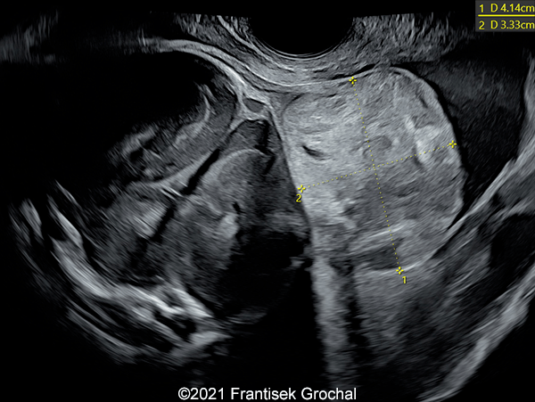
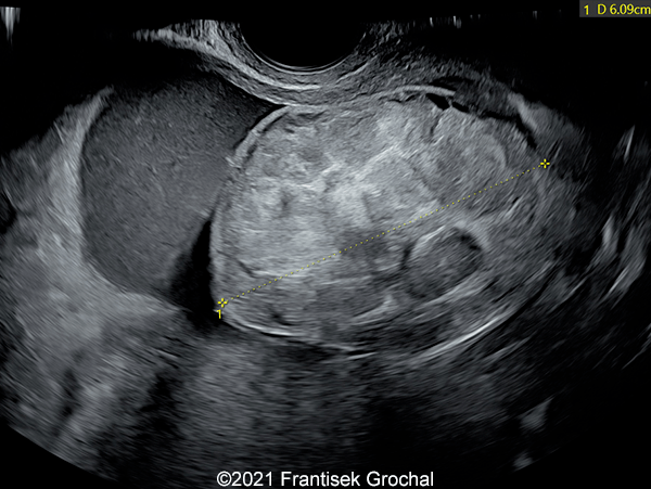
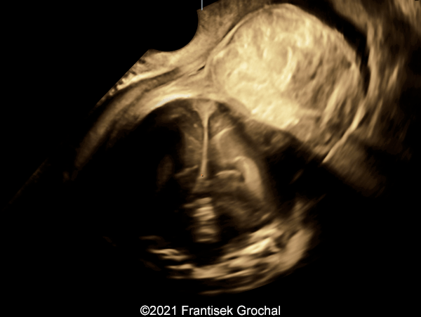
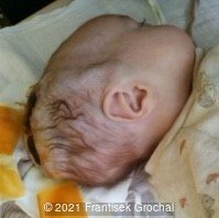
Case Report: A 24-year-old secundigravida, primipara presented to our office at 24 weeks and 6 days of her pregnancy due to a right-sided cranial parieto-occipital mass of the fetus. We obtained the following images.
View the Answer Hide the Answer
Answer
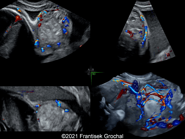
We present a case of hemangioma-hemangioendothelioma.
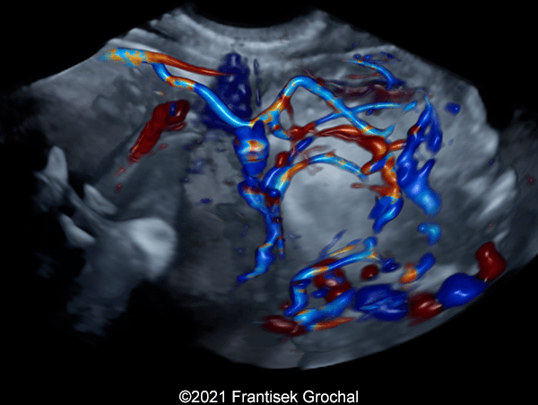
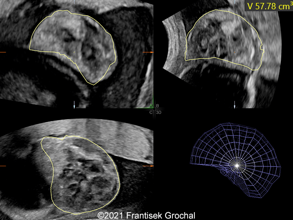
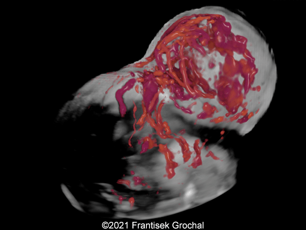
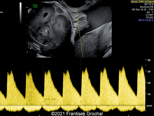
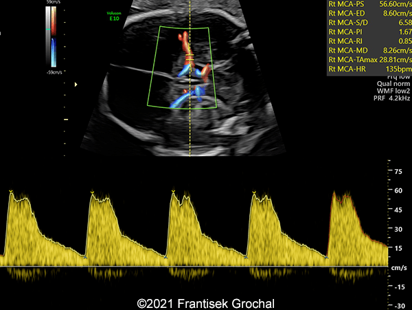
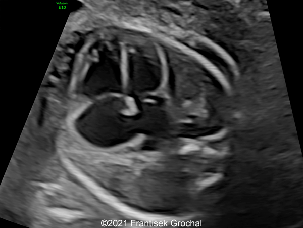
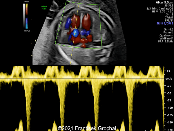
Our ultrasound images at 24 weeks, 6 days show a highly vascularized, mostly solid mass measuring 58 x 45 x 34mm in the right-parieto-occipital subcutaneous region. Vascular supply of the lesion could be traced to the right vertebral artery. Repeat ultrasound exams done later in pregnancy demonstrated that the size of the mass remained stable with overall mass volume about 58 cm³.
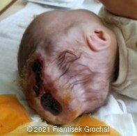
The mother was sent to The Institute for Mother and Child Care in Prague - Podolí, Czech Republic, for consultation and further management. A eutrophic male newborn was delivered via cesarean section at 38 weeks and 3 days due to non-progressive labor.
Diagnosis of hemangioma-hemangioendothelioma was clinically confirmed after delivery. The baby was placed on Propranolol treatment. At the time of posting this case, the baby is four months old and is doing well. The size of the tumorous mass diminishes gradually, and so no surgical treatment has been recommended thus far.
Discussion Board
Winners

Albert Buwono Indonesia Physician
Dianna Heidinger United States Sonographer
Javier Cortejoso Spain Physician

Seadet Zeynalova Azerbaijan Physician
Fatih ULUC Turkey Physician
Umber Agarwal United Kingdom Maternal Fetal Medicine
Igor Yarchuk United States Physician
Cem Sanhal Turkey Physician
Mayank Chowdhury India Physician
filiz halici öztürk Turkey Physician
Omayyah Dar Odeh Jordan Physician
Vladimir Lemaire United States Physician
DAVID BEAUMONT United Kingdom Physician
Ivan Ivanov Russian Federation Physician
Sara Abdallah Salem Egypt Physician
ELENA Novikova United States
Halil Mesut Turkey Physician
lan nguyen xuan Viet Nam Physician
Shafiga Hamzayeva Azerbaijan Physician
Andrii Telytchenko Ukraine Physician

Suat İnce Turkey Physician
Victoria Giang Viet Nam Physician
Shari Morgan United States Sonographer
KAORU YAMASHITA Japan Physician
Amparo Gimeno Spain Physician
Yasemin Dogan Turkey Physician
Ta Son Vo Viet Nam Physician
Selvanandhini Gopalasundaram India Physician
Murat Cagan Turkey Physician
Umutcan KAYIKÇI Turkey Physician
Sonio Sonio France AI
Rasha Abo Almagd Egypt Physician
DIMITRIOS SPILIOPOULOS United States
Sonia Roohollahi Iran, Islamic Republic of Radiologist
Ionut Valcea Romania Physician
alberto sosa olavarria Venezuela